COST course on Pharmaceutical Imaging in Lund, Sweden Back to the articles Back to the articles Sat November 3, 2012, 12:19 | Sat November 3, 2012, 12:19 |  13215x | 13215x |  Feel free to leave a comment Feel free to leave a commentTopic: Science | Series: My Journey to PhD 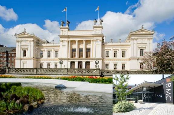 Recently I reported about COST supported course on the basics of Mass Spectrometry Imaging (MSI) that took place in Amsterdam at FOM-Institute AMOLF. At the end of October was organized a course for intermediate users of MSI in Bio Medical Center (BMC) of Lund University in Sweden. I took part together with two colleagues of mine from AMOLF and here I bring a summarizing report on the course. Recently I reported about COST supported course on the basics of Mass Spectrometry Imaging (MSI) that took place in Amsterdam at FOM-Institute AMOLF. At the end of October was organized a course for intermediate users of MSI in Bio Medical Center (BMC) of Lund University in Sweden. I took part together with two colleagues of mine from AMOLF and here I bring a summarizing report on the course. The course was called Applied MSI Pathology and Pharmaceutical Imaging. Its aim, from the educative point of view, was to give the students background about pathology and medicinal chemistry and to expose them to histology and pharma driven workflows of MSI. At AMOLF we are interested in biomolecules and biomarkers discovery rather than small molecules and quantification, so we decided to take the advantage of COST support and learn couple of new approaches and applications. I was really excited to connect two different parts of my work: pharmacy & imaging! Because COST is an international organization, the other purpose was to share the experience from laboratories across Europe. There were 13 students coming from 10 different countries having varieties of scientific backgrounds. I think we can all agree that the plan to share and exchange experiences was completely fulfilled. During the whole course we had lots of discussions on different topics and we had the opportunity to learn about various research projects and work groups. And beside we actually learned a lot about our cultures and enriched our word power in couple of languages. :-) To compare with the previous course in Amsterdam, the group felt much closer. What regards the educative aspects, I would however appreciate the course at AMOLF more, for mainly two reasons: 1/ despite the intermediate level of Lund course, many of the lectures were at a somewhat basic level. For me it was still quite fine, as I am still new to the topic, but for someone dealing with imaging for couple of years it could have been really a way too easy, 2/ we did not have much opportunity to perform our own experiments and most of the time during the afternoons in the labs we were just watching someone. I might also add that lot of the software or instruments just did not work when we needed, but in a lab, you never know… :-) But there were also couple of things I fancied: I am glad I could see how imaging experiments are carried out in a clinical oriented lab. MSI is of course not the only one technique used in such a research and it demands a co-operation with other bioanalytical techniques such as liquid chromatography and immunohistochemistry. I appreciated all the talks with the expert from Thermo, who actually answered a lot of my questions regarding the instrumentation and principles of Orbitrap. I liked the various debates, which were sometimes quite passionate. Perhaps when we were discussing the term spatial resolution. Another topic of our debates was the matrix application and optimization its parameters (concentration, solution, application device, layer thickness, form of crystals etc). We were trying to find the optimal way of matrix application, which would be fast enough, would ensure a homogeneous layer of crystals and would be reproducible. Our conclusion was that an ideal device should be a combination of ImagePrep (which most of us use for matrix application) and an airbrush sprayer (used in BMC). I guess we all appreciated the demonstration of a Laser Capture Microdissection (LCM) device. This device/technique uses light microscopy in combination with a laser dissection (cutting) to separate subpopulations of cells from a tissue region. The cells of interest can be then harvested to give histologically pure enriched cell populations. LCM has not been used a lot in combination with MSI, but it is a powerful and helpful tool in molecular analysis. To summarize, I learned couple of new MSI related points, I could compare work approaches in different work groups and I got to know bunch of great people from all over Europe. Thank you guys for nice time. :-) No comments. |
|
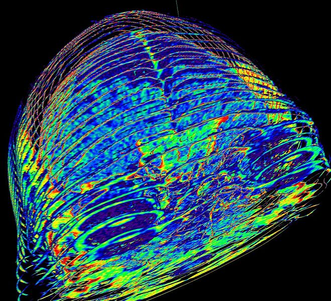
Photo No. 77: 3D CT scan of brain (Open in new window), Author: , Date: January 27, 2014
You are here: Karolinas.net » Home » » COST course on Pharmaceutical Imaging in Lund, Sweden
 Česky
Česky English