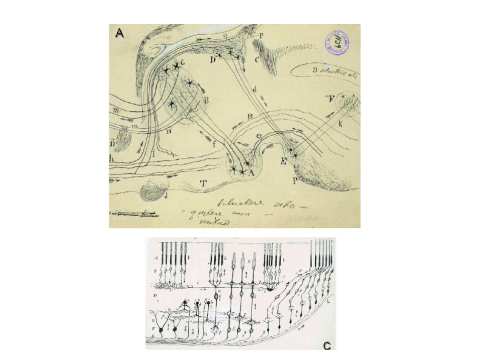
The brain research is IN more than ever before: BRAINPATH - Molecular Imaging of Brain Pathophysiology Back to the articles Back to the articles Sun January 26, 2014, 15:29 | Sun January 26, 2014, 15:29 |  23295x | 23295x |  Comments (1x) Comments (1x)Topic: Science | Series: My Journey to PhD 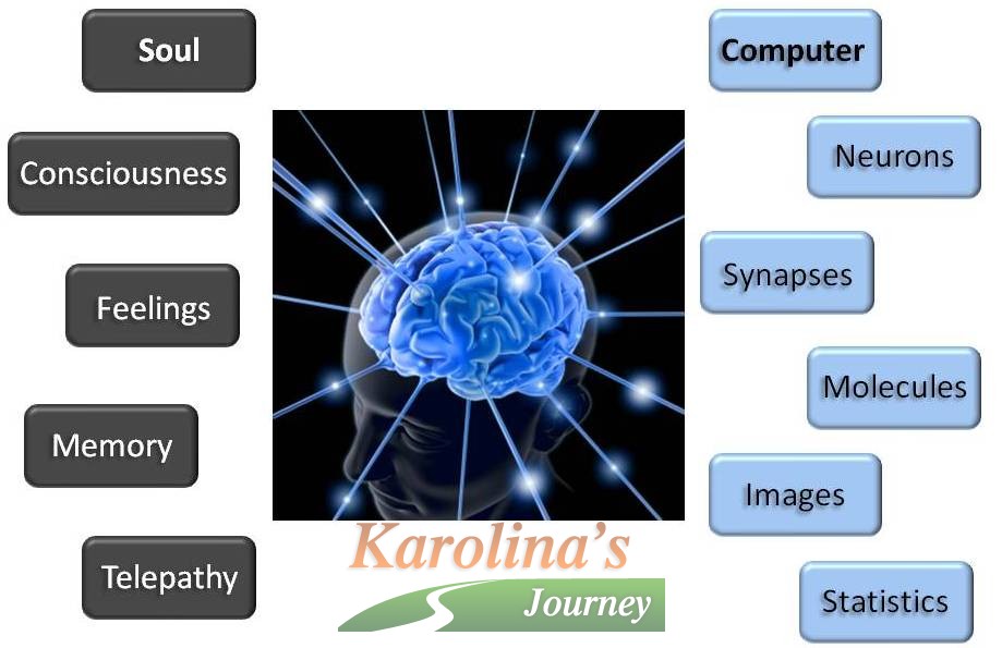 No other organ has excited the interest of scientists and philosophers as much as the brain. The long history of the brain research passed through many stages with different beliefs and views on what brain actually is, how it looks like, and how it works. The brain is at present understood as a biological computer, which operates and controls the whole body and the processes within. The brain research still attracts enormous attention. Desire to construct a computer with the capabilities of the human brain is strongly accented. Other research aims at brain pathophysiology and understanding of a mechanism of various nervous tissue related diseases. The latter takes the advantage of different imaging and scanning techniques which are capable to display distinct physiological and anatomical features of the brain. No other organ has excited the interest of scientists and philosophers as much as the brain. The long history of the brain research passed through many stages with different beliefs and views on what brain actually is, how it looks like, and how it works. The brain is at present understood as a biological computer, which operates and controls the whole body and the processes within. The brain research still attracts enormous attention. Desire to construct a computer with the capabilities of the human brain is strongly accented. Other research aims at brain pathophysiology and understanding of a mechanism of various nervous tissue related diseases. The latter takes the advantage of different imaging and scanning techniques which are capable to display distinct physiological and anatomical features of the brain. Before the brain research reached its current level, this organ was attributed different functions. For instance, Aristotle (4th century BC) believed the heart is the organ of thought and sensation and he considered the brain an organ designed for cooling the heart down. Aristotle, however, claimed that the organ of thought is not the same as the basis for thought. The basis for thought, as he described, is immaterial and cannot be found anywhere within the body [1,2,3]. Jumping in a time to the 17th century, we find Rene Descartes proposing that brain works as a machine. He described the functioning of the brain by the principle of a hydraulic pump coordinating the movements of the brain fluids. However, he concluded that this mechanism cannot account for some of the higher mental faculties found in man such as intellect and emotion. Instead he came up with the idea of a dualistic system in which the organ of the brain is distinguished from the immaterial "mind". In his view, it is the mind/soul, not the brain, which contains a person's thoughts, desires, and emotions [1]. The speculations about what the soul is, do we have one, and if yes where is it located, and is the soul equivalent to the brain have been living up to now, and have raised up many discussions. Whereas the materialistic approach rather disagrees with the soul’s existence and understands the brain fully as the seat of thought and emotions, other approaches try to grasp the essence of the soul. This is where "science" comes into contradiction with "pseudoscience", such as psychology, and mostly also with the religion. I am staying actually very open-minded when it comes to such discussions. I do not think that either the "real" science or the spirituality solely can explain the miracle of life. The two approaches should not been in opposition, but rather should take the most of each other. The brain research has even gained on importance in the past years. No wonder that many brain related scientific projects are under way. A few months ago I came across a short video about a project that took my full attention: The Human Brain Project. It is a "ten-year, large-scale European research initiative with the goal to understand the human brain and its diseases and ultimately to emulate its computational capabilities." The project seemed so cool to me that I wished to be part of it. Ten years to go… still a chance to get there after my PhD, I thought. However, before that could even happen I got a chance to be part of another brain research related project: the BRAINPATH. BRAINPATH employs the-state-of-art molecular imaging to understand the brain pathophysiology. It is a joint European initiative of three countries (The Netherlands, Belgium and Germany); eight research teams with the platforms of various medical imaging techniques are involved; seven work packages aim on the investigation of distinct pathological states/diseases, such as Alzheimer’s disease or glioblastoma, and on the aspects of the molecular imaging. The history of the brain imaging is more than a century long. One of the first imaging of brain tissue was performed using staining protocols, microscope, a pen and a sheet of paper. Santiago Ramón y Cajal and Camillo Golgi won the shared Nobel Prize for their work on structure and function of nerve cells in 1906 using this approach. While the latter one came up with the staining protocol, it was mostly Cajal, who used it in practice. The two of them were actually huge rivals [4].
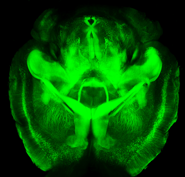
As the very first neuroimaging technique is actually considered the so called "human circulation balance" used by an Italian physiologist Angelo Mosso. The experiment seems to be rather funny nowadays. However, as other modern imaging techniques such as functional magnetic resonance imaging or positron emission tomography, Mosso’s technique was used to monitor the blood flow in working and resting states, both in physiological and pathological conditions [5]. Within BRAINPATH an orchestra of imaging techniques (A, B, C) is used to investigate the brain pathophysiology. The modern techniques, that are employed in medical practice, include magnetic resonance imaging (MRI), fluorescence imaging (FI), X-ray tomography & computed tomography (CT), positron emission tomography (PET), multi-spectral optoacoustic tomography (MSOT), and mass spectrometry imaging (MSI). The latter technique is the one that I am involved in. MSI maps the molecular distribution within the brain sections. It is an ex vivo technique, which means we do not work with living animals. The advantages of MSI lay particularly in its ability to not only localize but also to identify many different molecules from a brain section without a need for any labels, i.e. without the need to mark the to-be-analyzed molecules before the actual analysis. It can work in a discovery mode as well as it can serve for tracking down a particular compound within the tissue. Each of the imaging modalities offers several benefits, and simultaneously suffers from a few disadvantages. There is nothing like an ideal imaging technique capable of displaying every single aspect of the brain. Hence, a call for multimodal imaging is emphasized. Finding a way how to combine the imaging approaches is one of the ultimate goal of the BRAINPATH initiative. Several of the groups involved offer expertise in programming algorithms that can be used for a coregistration of the images acquired with different modalities. By such a combination a unique set of information about the brain and its diseases can be restored. Another challenge in brain imaging is the creation of the E-biobanks, i.e. databases which would contain all information about a particular pathological state of brain. At AMOLF, we are for instance involved in a working package of the COMMIT programme. "This project aims to establish an environment to process and evaluate large datasets originating from molecular histology using imaging mass spectrometry." A multimodal database would ideally include the information on the molecular composition acquired by mass spectrometry imaging, functional processes recorded by positron emission tomography and magnetic resonance imaging, contrast images from computed tomography scans, and possibly other. Examples of such biobanks already exist. For example the Allen Brain Atlas is a remarkable collection of brain sections images of gene expression. BRAINPATH and many other brain research related projects are unique initiatives that will help us to understand how our brain works and what happens if it does not work properly. To stay open-minded, we should not, however, forget that there might be something that cannot be measured by any of the current techniques. Even though there have been for instance very recently published two papers on fluorescent imaging of memory flow in special mouse models and in murine nervous cell cultures [6,7], it offers only the materialistic insight in our brains; we are stiil looking "only" at the molecules. An amazing article about new developments in brain imaging has been recently published in National Geographic.
3D reconstruction of the brain and eyes from CT scans
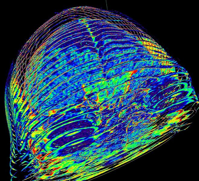
Brain is believed to have a grid-like structure.
[1] http://www.pbs.org/wnet/brain/history/index.html [2] http://www.mybrain.co.uk/public/learn_history1.php [3] C.G. Gross: Aristotle on the brain. The Neuroscientist 1 (1995) p245. [4] J.A. De Carlos, J. Borrell: A historical reflection of the contributions of Cajal and Golgi to the foundations of neuroscience. Brain Research Reviews 55 (2007) p8. [5] S. Sandrone, M. Bacigaluppi, M.R. Galloni, S.F. Cappa, A. Moro, M. Catani, M. Filippi, N.M. Monti, D. Perani, G. Martino: Weighing brain activity with the balance: Angelo Mosso's original manuscripts come to light. Brain (2013) [Epub ahead of print] [6] A.R. Buxbaum, B. Wu, R.H. Singer: Single β-Actin mRNA Detection in Neurons Reveals a Mechanism for Regulating Its Translatability, Science 343 (2014) p419. [7] H.Y. Park, H. Lim, Y.J. Yoon, A. Follenzi, C. Nwokafor, M. Lopez-Jones, X. Meng, R.H. Singer: Visualization of Dynamics of Single Endogenous mRNA as Labeled in Live Mouse. Science 343 (2014) p422. January 29, 2014, 00:19 (No. 130)Tom
Pěkný článek. Oceňuji "open mind" přístup (viz mozek a duše) |
|
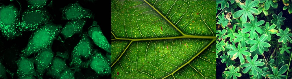
Photo No. 98: ImPressed by Brains, Plants and Molecules (Open in new window), Author: , Date: November 30, 2014
You are here: Karolinas.net » Home » » The brain research is IN more than ever before: BRAINPATH - Molecular Imaging of Brain Pathophysiology


 Česky
Česky English